|
|
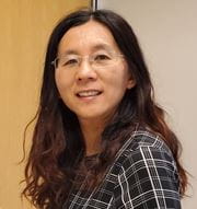
Hongli Yang, Ph.D.Research Scientist Phone: 503-413-5353 | Email: hyang@deverseye.org CV (Updated Nov 2024) | Peer Reviewed Publications |
|
Short Bio:
Dr. Hongli Yang graduated from Tulane University with a major in Biomedical Engineering and is a Research Scientist at the Optic Nerve Head Research Laboratory (ONHRL), part of Legacy Devers Eye Institute, Discoveries in Sight, at Legacy Research Institute. |
Publication Highlights:
OCT Optic Nerve Head Morphology in Myopia II: Peri-Neural Canal Scleral Bowing and Choroidal Thickness in High Myopia-An American Ophthalmological Society Thesis.
Diagnostic Performance for Detection of Glaucomatous Structural Damage Using Pixelwise Analysis of Retinal Thickness Measurements.
Optical Coherence Tomography Structural Abnormality Detection in Glaucoma Using Topographically Correspondent Rim and Retinal Nerve Fiber Layer Criteria. 3D Histomorphometric Reconstruction and Quantification of the Optic Nerve Head Connective Tissues. The Connective Tissue Phenotype of Glaucomatous Cupping in the Monkey Eye - Clinical and Research Implications. |
|
Research Interests:
|
||
Research Focus:
Dr. Yang has been working at the Devers Eye Institute Optic Nerve Head Research Laboratory (ONHRL), which is funded to study the effects of aging, glaucoma, and myopia on the neural and connective tissues of the optic nerve head using 3D histomorphometric reconstructions. This research has now expanded to include the use of Optical Coherence Tomography (OCT) to phenotype the optic nerve head, macular tissues, and peripapillary sclera in both monkeys and humans. This work has significantly contributed to a paradigm shift in how human patients with glaucoma or those at risk for developing the disease are imaged using OCT. 
Juvenile Myopia Research Project Looking for Participants:WHAT: Participate in the study Myopia related ONH Progression in Children, Teens, and Young Adults to help us learn about and better understand the features of myopia to help patients in the future. Participants will be evaluated every six months over an 18-month period, totaling four exams using clinical imaging devices. Exam time is 1.5-2 hours. REQUIREMENTS: Ages 6-22 with nearsightedness, ranging from-2D to -0.5D, family history of myopia, or axial lengths at an age at risk of developing myopia with a 50% chance or more. COMPENSATION: $50 per exam after completion. Primary Contact to sign up: myopiastudy@deverseye.org This study has been approved by the Legacy Institutional Review Board at Legacy Research Institute. High Myopia and Glaucoma Research Project Looking for Participants (all ages):Primary Contact to sign up: Cindy Albert, calbert@deverseye.org, 503-413-6457
|
||
MyHealth
MyHealth
Manage your account, request prescriptions, set up appointments & more.
Don't have an account
CREATE AN ACCOUNT >


