|
|
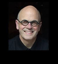
Claude F. Burgoyne, M.D.
Emeritus Van Buskirk Chair for Ophthalmic Research Email: cfburgoyne@deverseye.org
CV (updated Dec 2024) | Peer Reviewed Publications (Also see CV) |
|
Short Bio:Claude Burgoyne, MD is a retired Glaucoma Clinician Scientist and Emeritus Van Buskirk Chair for Ophthalmic Research at the Legacy Devers Eye Institute in Portland, Oregon. After an undergraduate Bachelor of Arts degree in Architecture and Medical School at the University of Minnesota, he pursued Ophthalmology residency training at the University of Pittsburgh and Glaucoma Fellowship training at the Wilmer Eye Institute of the Johns Hopkins Hospitals in Baltimore, MD. For twelve years he was Director of Glaucoma Services at the LSU Eye Center in New Orleans before moving to Devers in 2005. At Devers, he was Director of the Optic Nerve Head Research Laboratory (ONHRL) from 2005 to 2023. For 25 years his laboratory was NIH funded to study the effects of aging and experimental glaucoma on the neural and connective tissues of the monkey optic nerve head within 3D histomorphometric reconstructions. This work gradually expanded to include the cell biology of connective tissue remodeling and axonal insult early in the disease. Most recently, building upon its 3D visualization capabilities, his laboratory used Optical Coherence Tomography (OCT) to structurally phenotype the deep tissues of the human optic nerve head and peri-neural canal sclera in aging, glaucoma and myopia. Dr. Burgoyne's honors include the International Glaucoma Review AIGS Award (2004), the Lewis Rudin Glaucoma Prize (2008), the Alcon Research Institute Award (2010); the American Glaucoma Society 2015 Clinician Scientist Lectureship and the American Academy of Ophthalmology Achievement Award. He is currently an Emeritus member of the Glaucoma Research Society for which he will give the Goldmann Lecture at its annual meeting in November of 2024. He was inducted into the American Ophthalmological Society in 2023 and is a Gold Fellow of the Association for Research in Vision and Ophthalmology, (ARVO), for which he previously served as Glaucoma Section Trustee and President. |
Publication Highlights:The optic nerve head as a biomechanical structure: a new paradigm for understanding the role of IOP-related stress and strain in the pathophysiology of glaucomatous optic nerve head damage. The connective tissue phenotype of glaucomatous cupping in the monkey eye - Clinical and research implications. A biomechanical paradigm for axonal insult within the optic nerve head in aging and glaucoma. The morphological difference between glaucoma and other optic neuropathies. OCT Optic Nerve Head Morphology in Myopia II: Peri-Neural Canal Scleral Bowing and Choroidal Thickness in High Myopia-An American Ophthalmological Society Thesis. |
|
Research Interests:
|
||
MyHealth
MyHealth
Manage your account, request prescriptions, set up appointments & more.
Don't have an account
CREATE AN ACCOUNT >


 https://orcid.org/0000-0002-2765-4739